Our Equipment’s
First Point Dental modern clinics are equipped with the latest technology and newest equipments and state-of-the-art infrastructure to deal with your dental or oral healthcare concerns. Our dental team is quite adept at using advanced technology and equipment for our valued patients. In our consultation, we will brief you about the facilities and services we offer to deliver pleasant dental health care. Here is a guide to some of the latest and advanced equipment available at our disposal and what it offers. We would love to sit down with you and discuss your oral healthcare concerns. We will address it comfortably and swiftly. All you have to do is to schedule a consultation here or office a call at 888-736-6430
I-CAT System
Unlike the conventional 2D X-rays, we have invested in the renowned I-Cat Cone Beam 3D Imaging System. This is the latest and advanced state-of-the-art technology helping our dentists gain insights on oral healthcare. With the technology at the hand, we get access to high-definition visualization of your teeth, soft tissue, bone, and jaws. We will be able to identify important dental findings like extra teeth, airways concerns, or impacted teeth which isn’t possible with the traditional 2D technology. We will deliver accurate, insightful, and quick diagnoses. This would help in developing a treatment plan.
Important Features of the I-CAT System
- A single 20-second scan with accurate and important information for a complete dental diagnosis
- Less radiation exposure than the conventional CT scanners
- Increased level of comfort as the patients will sit in an open space
- The information can be easily shared with patients and we can take the right decisions
Our offices in both Hoffman Estates and Westmont offer i-CAT 3D imaging to our orthodontic patients.
Come visit today and find out more about the advanced, patient-friendly dental experience. Don’t wait! Call us today to schedule an appointment with leading dental specialists in Hoffman and Estates and Westmont locations.
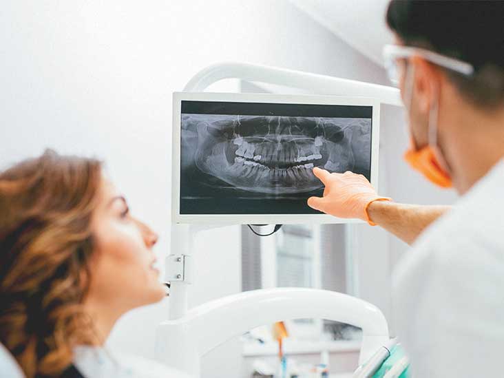
Orthopantomograph Machine
Orthopantomograph machine or OPG has reduced the treatment time at our dental offices. With the equipment, we will take a panoramic X-ray of your lower and upper jaws including the teeth. The machine will rotate around the patient’s head with the procedure taking just 20 seconds. The whole process is swift and painless with zero preparation time. Also, there is no radiation left in the body after the completion.
The advanced equipment comes in handy in cases like
- Dislocated Jaw
- Teeth Problems
- Gum Disease
- Mouth Infection
- Jawline Fractures in event of a trauma or accident
- Oral Surgery Planning
- Dental Implants
X-Ray Units in each Operatory
Giving utmost care to our dental patients lies at the core of our services. To do so, we have invested in top-of-the-line dental equipment with a specialized team of dentists at your disposal. A visual oral examination is incomplete without a dental x-ray machine. Essentially, X-Rays help us understand your jawbone’s strengths and weaknesses and its condition. We gather information and accurate insights into your oral health and helps us in identifying any possible risk of developing gum disease or tooth decay. Post the X-rays results, we can take preventive action before your oral health takes a turn for the worse.
Is it safe to undertake an X-Ray dental examination?

As we all know that the X-ray machine emits radiation but there is nothing to worry about for a bit. The devices are specifically customized and produced for safe dental care. It’s safe for both the patients and dentists at our dental clinics. We follow the stringent ADA (American Dental Association) regulations regarding X-Ray use. We advise our patients to wear protective aprons over their collar and abdomen for thyroid protection. If you are pregnant or lactating, do intimately with your dentist beforehand. This will help the dentist in identifying if X-rays are necessary or not. If not, it will be performed at a later time.
Mouthwatch Intraoral Camera
We are able to examine the patient’s inner mouth accurately with the help of captured digital images. This helps us gain better insight into one’s oral health while building up case studied and patient file documentation. Usually, we will connect it to a computer wirelessly through a docking station or USB port. The device captures the images with the aid of high beam lights without the assistance of any external lighting. Our device can produce high-quality images in a short span of time.
